Running Head: Belimumab Reduces Autoantibodies in COPD
Funding Support: This study was supported by an investigator-initiated research award from GSK. The sponsor did not have a role in study design; in the collection, analysis, or interpretation of data; in the writing of the report; or in the decision to submit the report for publication.
Date of Acceptance: September 15, 2026 | Published Online Date: October 6, 2026
Citation: Wade RC, Robison S, Wells JM, et al. A phase 2b randomized trial of belimumab to reduce atherogenic autoantibodies in COPD patients.
Abbreviations: AE=adverse event; anti-GRP78=glucose-regulated protein 78; AST=aspartate aminotransferase; BLOCK-COPD study=Beta-Blockers for the Prevention of Acute Exacerbations of Chronic Obstructive Pulmonary Disease trial; CAC=coronary artery calcification; CAD=coronary artery disease; cIMT=carotid intima thickness; COPD=chronic obstructive pulmonary disease; CT=computed tomography; CTD=connective tissue disease; CVD=cardiovascular disease; DLCO=diffusing capacity for carbon monoxide; DSMB=data safety monitoring board; ELISA=enzyme-linked immunosorbent assay; FEV1=forced expiratory volume in 1 second; FVC=forced vital capacity; HLA-DR=human leukocyte antigen – DR isotype; HSP=heat shock protein; IgA=immunoglobulin A; IgG=immunoglobulin G; IL=interleukin; IRB=institutional review board; IV=intravenous; LAD=left anterior descending artery; LCX=left circumflex artery; LM=left main artery; OD=optical density; OR=odds ratio; PFT=pulmonary function test; PPA=polysaccharide antigens; PYs=pack years; RCA=right coronary artery; SAE=serious adverse events; SE=standard error; SEM=standard error of the mean; SLE=systemic lupus erythematosus; TNF-α=tumor necrosis factor-alpha; UAB=University of Alabama at Birmingham
Citation: Wade RC, Robison S, Wells JM, et al. A phase 2b randomized trial of belimumab to reduce atherogenic autoantibodies in COPD patients. Chronic Obstr Pulm Dis. 2026; 13(6): 440-451. doi: http://doi.org/10.15326/jcopdf.2026.0805
Introduction
Chronic obstructive pulmonary disease (COPD) is a leading cause of morbidity and mortality worldwide.1 COPD is most frequently caused in high-income countries by inhalation of tobacco smoke and/or other noxious fumes and is manifested by chronic airway inflammation and/or pulmonary parenchymal destruction that results in expiratory airflow obstruction.1,2
In addition to those lung injuries directly attributable to the toxic inhalations, COPD has many features of an autoimmune disease.3-6 Numerous lymphocyte processes that are pathognomonic for autoimmunity are common among COPD patients, including specific T-cell and B-cell autoreactivities to dozens of different self-proteins, and the magnitude of these abnormalities are often correlated with lung disease severity.3-8 Furthermore, some of the lymphocyte reactivities against autologous extrapulmonary proteins have been implicated in the pathogenesis of systemic comorbidities that afflict COPD patients.3,5,6
The most serious comorbidity of COPD is atherosclerotic cardiovascular disease (CVD), which is a major cause of death in this lung disease population.9-12 The increased frequency of CVD in COPD patients is not fully explained by other pro-atherogenic risk factors, including the extent of smoking, age, hypertension, diabetes, body habitus, race, educational level, or hypercholesterolemia.12 Moreover, the number and severities of serious complications following ischemic events is worse among COPD patients than in those who do not have this lung disease.9 The systemic inflammation that is frequently present in COPD patients is thought to possibly be a contributing factor for CVD,3,9-12 but mechanistic details of the causal immune process(es) have not yet been defined.
Immunoglobulin G (IgG) autoantibodies with specificities against glucose-regulated protein 78 (anti-GRP78) are prevalent in COPD patients.5,6 GRP78 is a heat shock protein (HSP) that has important functions as an intracellular protein chaperone and component of the unfolded protein response.13,14 In addition to its localization in cytoplasm and endoplasmic reticulum, GRP78 can also be facultatively secreted into extracellular spaces, where it binds to cognate cell surface receptors that transduce danger signals.13,14
The functional effects of GRP78 receptor engagements are magnified by cross-linking those complexes with multivalent (i.e., IgG) anti-GRP78 autoantibodies.5,6,13-15 Among other consequences, patient-derived anti-GRP78 IgG induces human monocytes, macrophages, and endothelial cells to increase their NFκB phosphorylation and productions of interleukin (IL)-8, C-C motif chemokine ligand 2 (monocyte chemoattractant protein-1), matrix metalloproteinase-9, tumor necrosis factor-alpha (TNF-α), interferon regulatory factor 7, and adhesion molecules, all of which have roles in atherogenesis.5,6,13-15 Like other IgG autoantibodies, anti-GPR78 can also readily fix complement and form antibody-antigen complexes: these highly cytotoxic processes also cause dysregulated proliferations of vascular smooth muscle and endothelial cells that promote atheroma maturation.16 Experimental in vivo generation of anti-GRP78 autoantibodies in susceptible mice, or adoptive transfers of these autoantibodies into the animals, recapitulates the development of arterial atherosclerosis.15
We have previously shown that circulating anti-GRP78 levels in COPD patients are highly associated with carotid intima thickness (cIMT) and, importantly, this relationship was independent of other known CVD risk factors, including plasma levels of TNF-α, IL-6, and C-reactive protein.6 Epitope discovery assays further showed the link to cIMT was strongest for the subset of anti-GRP78 autoantibodies with specificity for a linear, internal GRP78 peptide sequence of amino acids 246-260 (GRP78246-260).6 Anti-GRP78 autoimmunity has also been implicated in osteoporosis and emphysema in COPD5 and is common too among patients with connective tissue diseases (CTDs), which are similarly notable for frequent and severe CVD.13,14 Of importance, the CVD risk in CTD patients is reduced by treatments that specifically target their underlying autoimmune abnormalities.17-20
We hypothesized GRP78 autoreactivity may also be involved in the pathogenesis of coronary artery disease among COPD patients. We further speculated that mechanistically focused interventions that decrease anti-GRP78 IgG in this population could potentially ameliorate the frequency and/or severity of their CVD.17-20 The safety of autoantibody reduction in COPD patients, however, had not yet been tested. However, belimumab, a humanized monoclonal antibody inhibitor of B-cell activating factor, has a favorable safety profile when used as adjunct therapy in patients with active systemic lupus erythematosus (SLE), many of whom also have chronic lung disease.21,22
Accordingly, we examined the association of anti-GRP78 humoral responses with CVD in COPD patients, and then conducted a randomized, placebo-controlled trial to assess the feasibility of using belimumab for autoantibody reduction in this disease population.
Methods and Materials
Correlations of Anti- Glucose-Regulated Protein 78 Autoantibody Concentrations with Coronary Artery Calcium
Participants
The discovery cohort for the initial anti-GRP78 and coronary artery calcification (CAC) association analysis consisted of University of Alabama at Birmingham (UAB) participants in the Beta-Blockers for the Prevention of Acute Exacerbations of Chronic Obstructive Pulmonary Disease trial (BLOCK-COPD Study)23 who had prior noncardiac gated chest computer tomography (CT) scans and stored frozen plasma. The subsequent validation analysis was performed using identical materials from UAB participants in the Prospective Repository for coupling EVEnts to Novel paThways in COPD and Asthma (PREVENT) cohort who similarly had prior CT scans and residual plasma specimens. All participants had COPD associated with tobacco smoking, with fixed expiratory airflow obstruction on pulmonary function tests (PFTs), as shown by ratios of expiratory volume in the first second of a forced expiration (FEV1) to forced vital capacity (FVC) <0.7. Each participant also completed demographic and medical history questionnaires and provided written informed consent. These studies were approved by the institutional review board (IRB) at UAB (IRB 150609007, IRB-170301002).
Lung Function and Imaging Assessments
PFTs were performed using standard methodologies, and best postbronchodilator values were tabulated and analyzed. Chest CT scans were used to determine CAC lesions by Weston criteria.24 CT scans were interpreted and scored by 2 investigators blinded to the autoantibody assays. The Weston visual scoring system was used rather than the Agatston method because CT scans in both cohorts were noncardiac-gated and obtained for indications other than dedicated calcium scoring. This visual scoring approach assigns a severity grade of 0–3 to each of 4 coronary artery territories (right coronary artery, left main, left anterior descending, and left circumflex), for a total score ranging from 0–12, and has been shown to perform comparably to the Agatston score for prediction of prevalent coronary artery disease and incident cardiovascular events in smokers, including those with COPD, imaged with nongated CT.24
Glucose-Regulated Protein 78 Enzyme-Linked Immunosorbent Assay
Concentrations of circulating anti-GRP78 IgG autoantibodies were measured by enzyme-linked immunosorbent assay (ELISA), as detailed previously.6 In brief, these assays detect plasma autoantibodies with specificities for solid phase ligands consisting of either plate-bound intact, whole recombinant GRP78 or a synthetic peptide corresponding to GRP78 amino acids 246-260 (GRP78246-260). The latter was recently shown to be the epitope for anti-GRP78 autoantibodies with the strongest correlation to cIMT.6 The target peptide (246-ATNGDTHLGGEDFDQ-260) was biotinylated at the N-terminus after addition of an aminohexanoic linker to minimize steric hindrance and added to wells of a streptavidin-coated ELISA microplate. After incubations with diluted patient plasma, washes, and addition of detection reagent, as detailed previously,6 optical densities (OD) were determined at 405nm in triplicate measures of each specimen.
Belimumab Clinical Trial
Objectives
To our knowledge, therapy that specifically targets autoantibodies has never previously been tested in patients with COPD. Thus, this phase 2b double-blinded, placebo-controlled, randomized clinical trial was designed to be a pilot "proof-of-concept" study to establish feasibility and begin to explore efficacy and safety of belimumab treatment in COPD patients.
Participants
Trial participants were outpatient volunteers recruited from the UAB Lung Health Center who gave informed consent, in accordance with a protocol approved by the UAB IRB (IRB-300000119). The study was also overseen by an independent data safety monitoring board (DSMB).
Participant Enrollment Criteria
Inclusion criteria included a diagnosis of COPD (FEV1/FVC< 0.7, and FEV1 between 25% and 80% of predicted values) attributed to cigarette smoking, age 40–75 years old, and levels of plasma IgG autoantibodies against GRP78246-260 that were in the upper 3 quartiles of values seen in previously tested COPD patients (i.e., ≥0.178 OD units)
Exclusion criteria included a history of an acute COPD exacerbation requiring treatment within the preceding 12 months or any exacerbation within the last 4 months, current smoking or use of systemic steroids or cellular immunosuppressants within the preceding 6 months, a history, clinical, or laboratory evidence of other autoimmune syndromes, malignant neoplasm within the last 5 years, serological evidence of past/current hepatitis B or C, history of a primary immunodeficiency, or hospitalization for treatment of infection or use of a parenteral antimicrobial agent within 60 days of screening.
Randomization and Treatment Arms
Following successful screening, participants were randomized by computer in the UAB Investigative Pharmacy to experimental (belimumab) or control arms (2:1, respectively). Patients, staff, and investigators were blinded to these assignments. All trial medications were administered by Center staff.
1) Belimumab: A target of 12 experimental treatment participants were to receive 3 doses of intravenous (IV) belimumab (10mg/kg), each 2 weeks apart, followed by 5 more infusions at monthly intervals.
2) Placebo: 6 control participants were to receive identically appearing placebo IV on the same schedule as the belimumab individuals.
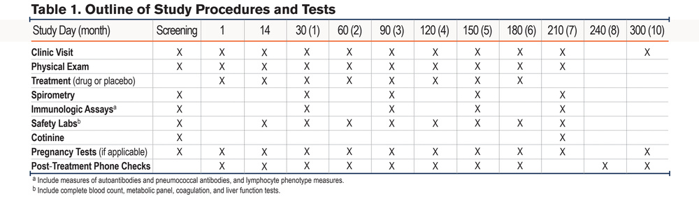
The schedule of trial procedures are also detailed in Table 1.
Endpoints
The primary endpoint was a determination of treatment effects on concentrations of circulating anti-GRP78246-260 autoantibodies, comparing plasma levels at baseline to those at the conclusion of therapy (or at last visit, if participation was terminated early). Secondary endpoints of the trial were: (1) treatment effects on concentrations of pneumococcal polysaccharide-binding antibodies, determined by ELISA; (2) treatment effects on proportions and phenotypes of circulating B-cells, ascertained by flow cytometry; and (3) comparisons of adverse events (AEs), occurring at any time during individual participation. National Cancer Institutes’ Common Terminology Criteria for Adverse Events scales were used to grade these events.
Pneumococcal Enzyme-Linked Immunosorbent Assay
Protective IgG antibodies specific for pneumococcal polysaccharide antigens (PPA) were measured by sandwich ELISA in plasma obtained from participants at baseline and their last study visit. Assays were performed using reagents and methods provided in a kit (Abnova; PCP IgG ELISA, #KA4831; Taipei, Taiwan). These ELISA measured aggregate IgG with specificity against 23 PPA serotypes (1-5, 6B, 7F, 8, 9N, 9V, 10A, 11A, 12F, 14, 15B, 17F, 18C, 19F, 19A, 20, 22F, 23F, 33F) and were denoted as binding units per ml (U/ml).
Flow Cytometry
Peripheral blood lymphocytes, immediately prior to and at the conclusion of therapy, were isolated from venipuncture specimens by density gradient centrifugation using reagents, methods, analyses, and equipment previously detailed.4-6,8
Other Data
Additional analyses that were not formal endpoints included assessments of treatment effects on spirometric values and circulating T-cell numbers and phenotypes ascertained by flow cytometry.4-6
Statistical Analyses
Unpaired continuous or ordinal variables (e.g., demographic parameters) were compared between treatment arms using the Kruskal-Wallis test. Categorical variables (e.g., genders) were compared by using Chi-square statistics. Spearman rank order correlations were used to identify associations between continuous and ordinal variables (e.g., the relationship between anti-GRP78 and CAC). Wilcoxon signed-rank tests were performed to compare paired continuous or ordinal variables (e.g., baseline and final anti-GRP78246-260). Poisson regression models were used to evaluate associations of CAC with anti-GRP78, adjusted for covariates of interest. Analyses were performed with StatView v5.0.1 (SAS Institute; Cary, North Carolina). Unless otherwise denoted, results are depicted as means ± standard error of the mean (SEM). Results were determined to be statistically significant when the accompanying test yielded a probability of <0.05.
Results
Glucose-Regulated Protein 78 Enzyme-Linked Immunosorbent Assay and Coronary Artery Calcium Associations
Participants
Characteristics of the discovery and validation populations for these analyses are detailed in Table 2. The 2 study cohorts were well matched with respect to age, sex, and smoke exposures. There was an insignificant trend for a larger proportion of White participants among the discovery cohort and COPD severity was also greater in that group. White and Black participants had similar overall FEV1: (1.28±0.52L versus 1.34±0.59L, respectively) and FEV1/FVC: 0.46±0.15L versus 0.52±0.12L, respectively).
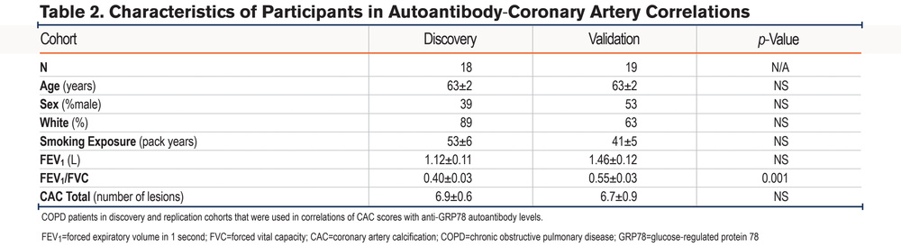
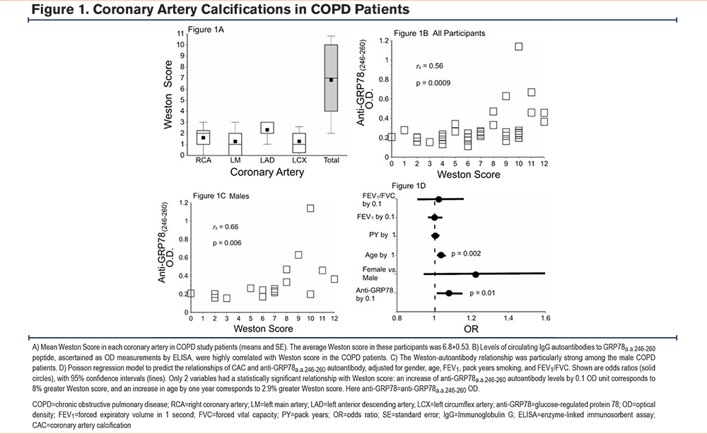
Weston scores were similar in both cohorts (Table 2), with observed values ranging from 0–12 across the combined cohort. There were similarly no intergroup differences between males and females (6.6±3.4 and 7.0±3.2 coronary calcifications, respectively) nor between White and Black participants (6.9±3.1 and 6.7±3.9 calcifications, respectively). There were no significant correlations between Weston scores and FEV1 (rs=0.09, p=0.59) or FEV1/FVC (rs=0.01, p=0.91). All but one COPD participant had at least one CAC abnormality. The anatomical distribution of the coronary artery lesions is depicted in Figure 1A.
Autoantibody Correlations With Coronary Artery Calcium
There was no significant relationship between the number of CAC abnormalities and ELISA values for circulating antibodies against full-length recombinant GRP78 within the discovery cohort (rs=0.18, p=0.45). However, and analogous to a previous finding linking anti-GRP78 with carotid artery disease in COPD,6 there was a much stronger correlation between the CAC abnormalities and autoantibodies with specificities for an internal GRP78 epitope (anti-GRP78246-260) (rs=0.67, p=0.006).
Subsequent study of the validation cohort again found anti-GRP78246-260 and CAC were related (rs=0.55, p=0.019). The correlation of anti-GRP78246-260 and CAC within the aggregate population (combined discovery and validation cohorts) is depicted in Figure 1B.
As noted in the study of autoimmune carotid artery disease in COPD,6 the correlation between anti-GRP78246-260 and atherosclerosis tended to be stronger among males (n=17) (Figure 1C), compared to the 20 females (rs=0.44, p=0.06). Anti-GRP78246-260 levels were not associated with baseline FEV1 (rs=-0.18, p=0.91) or FEV1/FVC (rs=0.009, p=0.96) of the participants but were weakly related to their ages (rs=0.39, p=0.02).
The increased risk of CAC abnormalities associated with anti-GRP78 was determined by estimating Poisson regression models with and without covariates. Unadjusted odds ratio estimates reveal that an increase in anti-GRP78 IgG by 0.1 OD units corresponded to a 9% increase in CAC. After adjustment by spirometric indices, sex, age, and smoking history, anti-GRP78246-260 autoantibody level increases by 0.1 OD unit corresponded to 8% greater Weston scores (Figure 1D).
Belimumab Clinical Trial
Participants
Recruitment ended prior to fully reaching targeted enrollment, and some experimental treatments and assessments were terminated early by patients due to their safety concerns (and/or institutional prohibitions) during the COVID-19 pandemic (Figure 2). Seventeen participants were randomized (belimumab n=11; placebo n=6) and participated in the trial for varying durations between August 9, 2018 and December 12, 2021. One participant was diagnosed with a gastric carcinoma soon after randomization and was withdrawn from further study; this serious adverse event (SAE) was believed to be unrelated to the experimental intervention by both the investigative team and the DSMB. This SAE was also adjudicated to be a likely confounder (given dysregulated constitutive productions of anti-GRP78 autoantibodies by carcinomas)14 and this individual was not included in the final analyses.
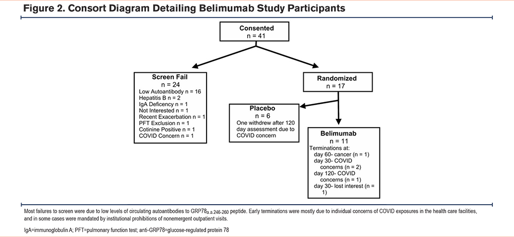
A total of 16 other participants participated for at least one month and were evaluated: 10 in the belimumab arm and 6 placebo controls (Figure 2). FEV1, as percentages of predicted values, ranged from 27% to 77% and, like other demographic or clinical parameters, were comparable in both treatment arms (Table 3).
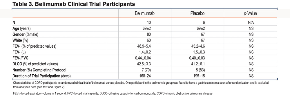
Given the small trial populations, with several early terminations and/or missing interval assessments, analyses were limited to comparisons of baseline (pretreatment) values with those measured at the participant’s last clinic visit.
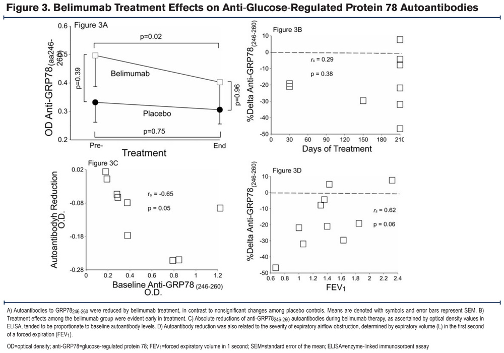
Belimumab treatments resulted in reductions of plasma anti-GRP78246-260 from baseline (pretreatment) values of 0.50+0.34 OD units to 0.40+0.09 OD units in the last available specimens (p=0.02), a decrement of 17+5%. In contrast, plasma autoantibodies among placebo controls did not appreciably change (p=0.75) (Figure 3A). Treatment effects were not obviously dependent on duration of therapy, even among participants whose treatments were terminated early (Figure 3B), but were highly associated with baseline concentrations of the autoantibody: anti-GRP78246-260 reduction was proportionately greatest among those with the highest initial autoantibody levels (Figure 3C). Treatment effects also tended to be proportionate to the severity of expiratory airflow obstruction immediately prior to beginning therapy (Figure 3D).
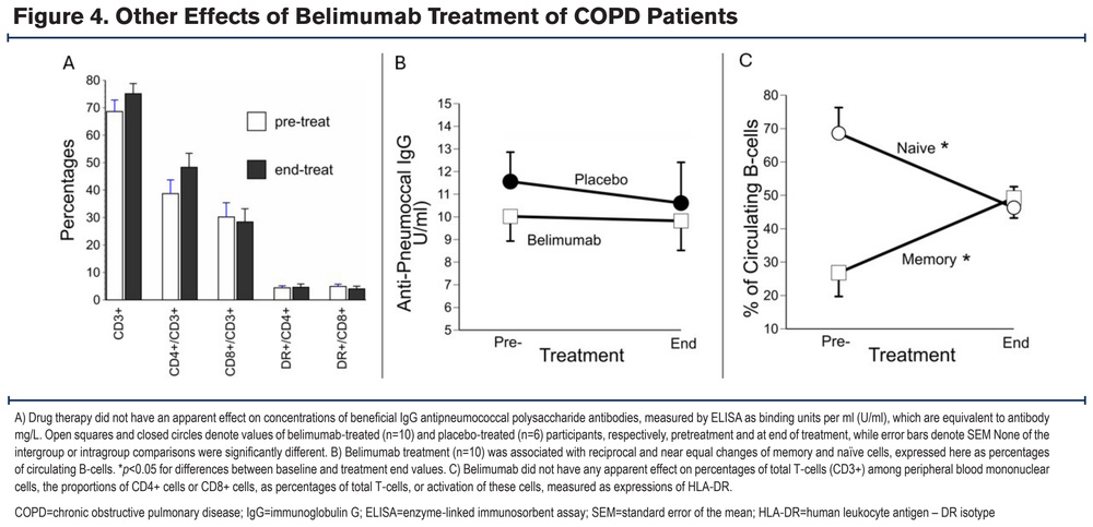
Levels of pre-existent “protective” (and highly beneficial) antibodies against pneumococcal antigens were not significantly altered by belimumab treatment (Figure 4A). CD20+ B-cells, as percentages of peripheral blood mononuclear cells immediately prior to therapy (4.3±0.7%), were not significantly different at the conclusion of the belimumab treatment (3.2±0.4%). However, drug treatment was associated with a nonsignificant decrease of transitional B-cells (from 5.5±0.8% to 2.8±0.8% of total B-cells), as well as significant reductions of the naïve subset, and reciprocal increases in the proportions of memory phenotypes (Figure 4B). None of these cell types varied in the placebo participants by >2% over the course of this trial (data not shown). T-cell numbers and phenotypes were unchanged by belimumab therapy (Figure 4C). Pretreatment and end-of-treatment FEV1 did not significantly differ in the belimumab group (1.39±0.16L and 1.28±0.17L, respectively), and FEV1/FVC was similarly unaltered by the experimental therapy (0.43±0.04 versus 0.40±0.04, respectively).
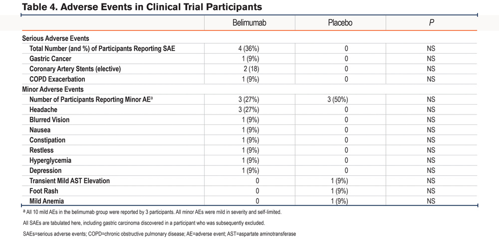
A total of 4 SAEs occurred in the belimumab group (Table 4). With the possible exception of a single COPD exacerbation, however, none of the SAEs were adjudicated to be a likely result of the intervention (Table 4). Several self-limited AEs of lesser severity occurred in both trial arms (Table 4).
Discussion
The initial correlative study described here showed a significant association between CAC scores and levels of IgG autoantibodies specific for an internal, linear GRP78 peptide epitope (anti-GRP78246-260) among 2 distinct cohorts of COPD patients (Figures 1B and 1C). As such, these data extend and substantiate a previous finding that circulating anti-GRP78246-260 autoantibodies were also highly correlated with the severity of carotid artery abnormalities in COPD patients.6 To our knowledge, the present study is the first report that links an antigen-specific autoimmune response to CVD in COPD patients.
The mechanisms by which autoimmune responses, including anti-GRP78, develop in COPD is currently unknown, but inferences can be drawn from findings in other, more thoroughly studied autoimmune syndromes. Adaptive immune responses that are initially directed at “appropriate” antigens (e.g., determinants of pathogenic microbes) can generalize or spread under certain circumstances to inappropriately target bystander self-proteins, and in many cases, these dysfunctional secondary autoimmune responses are far more injurious than the inciting infection.25,26 Secondary autoimmune responses due to epitope spreading are particularly prone to target self-proteins that are in overabundance proximate to the initial infectious or inflammatory foci.27-29 HSP production is upregulated by various stressors, such as exposures to noxious fumes and gasses, mechanical tension, infections, inflammation, and hypoxemia; and expression of GRP78 is markedly increased in the lungs of smokers and COPD patients.5,30 The development of autoimmunity is also promoted by the presence of sequence or conformational similarities between antigens expressed by an infective organism and the orthologous self-protein(s) of the human host (i.e., epitope mimicry).31,32 Many HSPs (including GRP78) are highly conserved throughout phylogeny, from microbe to man.33 In addition, highly reactive chemicals present in tobacco smoke can covalently modify native proteins to the extent they become neoantigens, for which immune tolerance had never developed, and these too are capable of evoking deleterious autoimmune responses.34
By whatever means they arise, secondary autoimmune responses tend to be chronic and/or recurrent, since the self-proteins that drive these responses are necessarily present indefinitely and/or are continually produced/renewed.3 This paradigm could perhaps account, at least in part, for observations that many COPD manifestations (including CVD) typically, if otherwise inexplicably, persist and often even progress, despite removal of the inciting agent (e.g., after smoking cessation). The persistence of self-antigens and consequent chronicity of many autoimmune responses could also contribute to the increased severity of late-occurring complications of CVD in COPD patients, such as mortality, heart failure, and arrhythmias.9
The possibility that targeting the underlying autoimmune process with a specific disease-modulating therapy might have a beneficial effect on CVD in COPD patients, as it does in other autoimmune syndromes,18-20 led us to initiate the belimumab trial. This study was intended to explore the feasibility of autoantibody reduction in COPD, while limiting patient risks by use of a small number of participants for a relatively short duration, while focusing on a rapidly responding biomarker (anti-GRP78 IgG) as the primary endpoint. This conservative trial design was prompted by concerns that the microbial colonization that is frequent, if not ubiquitous, in COPD lungs,35 might progress to more fulminant infections if host immune defenses were critically impaired by the belimumab.
Nonetheless, belimumab has a favorable safety profile in SLE patients, who not infrequently also have severe lung disease (and are, thus, also at increased risk of chronic/recurrent lower respiratory tract microbial colonization).21,22 Moreover, current prescribing recommendations for belimumab urge caution when treating patients who are known to have serious or chronic infections but fall short of delineating these comorbidities as absolute contraindications.36 Belimumab has disproportionately greater effects on less mature B-cell subsets: treated patients typically exhibit reductions of naïve and transitional cells, while long-lived memory B-cells are less affected and/or increase as a proportion of the total B-cell compartment,21,22 as was seen here (Figure 4B). Even though belimumab rapidly reduces levels of deleterious autoantibodies, titers of long-lived, protective antibodies against pneumococcus, tetanus, and influenza (and presumably other pathogens) tend to be maintained, as indicated in the present study (Figure 4A), a putative consequence of a relatively preserved memory cell compartment.21,37
In other indications, prolonging the duration of belimumab treatment results in progressively greater reductions of disease-associated autoantibodies, and greater clinical benefits.38-40 The ~17% reduction of anti-GRP78 aa246-2+0 here is comparable in magnitude to anti-double stranded DNA decreases after similarly short-term belimumab trials in SLE patients. Anti-dsDNA autoantibody levels are further reduced in those SLE trials after one year of belimumab,38-40 leading us to hypothesize that more prolonged drug treatment than we employed here might similarly result in additional reductions of anti-GRP78246-260 levels in COPD patients with CVD.
We believe the present study is the first systematic trial of targeted autoantibody reduction in COPD patients. These belimumab treatments decreased levels of a pathogenic autoantibody known to deleteriously affect many biologically relevant functions, recapitulate atheroma generation in vivo, and is correlated with the severities of carotid atherosclerosis in COPD cohorts.5,6,15 Belimumab therapy in the COPD participants here was also associated with preservation of antibodies important for host defense (Figure 4A) and minimal attributable SAE. Headaches frequently occur with monoclonal antibody therapy, and it is likely that those events reported here were due to the belimumab (Table 4), but those side-effects were mild and self-limited. It is also possible that the acute disease exacerbation in one belimumab patient (which resolved without sequela) was due somehow to the drug (Table 4), but these events are common in COPD patients, and this single case does not allow a meaningful analysis.
While these pilot trial results are encouraging, we also note this study has limitations. On the one hand, finding this intervention had the desired effects (Figure 3A), despite small numbers of participants and short (and often abbreviated) treatment courses, attests to the efficacy of the drug in this population. Longer treatment in more participants might demonstrate even greater drug effects21,38,39 or, on the other hand, result in a greater number or severity of adverse events. Atherosclerosis is a complex syndrome and, as such, other immune mechanisms that were not studied here may also contribute to CVD genesis in COPD.9-12,40 Another potential limitation is that BLOCK-COPD excluded patients with an established indication for beta-blocker therapy, though coronary artery disease (CAD) remained common in this cohort (15% history of CAD; 84.4% of an imaging subset had Weston score >0).23,41 This approach may limit generalizability to the full spectrum of CVD severity in COPD. Next, our use of the visual Weston score rather than automated Agatston quantification reflects the nongated acquisition of the CT scans in both cohorts; while Weston scoring is more subjective than automated methods, it has been validated for this purpose and performs comparably to Agatston scoring for prediction of CVD outcomes in COPD populations.42 Future studies incorporating dedicated, electrocardiogram-gated CAC scoring could provide more granular quantification of calcium burden. Most importantly, while belimumab reduced the concentrations of anti-GRP78, a singular autoantibody strongly implicated in atherogenesis and other COPD comorbidities,5,6,15 the present data cannot prove that these autoantibody reductions will result in detectible clinical benefits in this population. The extent to which progressive CVD can be ameliorated and/or over what time frame this might occur remains uncertain. To detect a favorable treatment effect on CVD progression would likely require precise serial measures in large numbers of participants over prolonged intervals. Alternatively, use of biologically relevant surrogate CVD markers, such as measures of arterial artery stiffness,43 might be a practical endpoint for subsequent clinical trials.
Given the high toll of mortality, disability, and health care expenditures caused by CVD among COPD patients, and the limitations of conventional treatments, considerations of novel therapies seem well justified. The studies presented here lend further credence to the paradigm that autoimmunity can play an important role in the pathogenesis of atherosclerosis and, moreover, also illustrate an innovative treatment approach that merits further evaluation in COPD patients with CVD.
Acknowledgement
Author contributions: RCW, SR, JMW, TM, YK, MTD, and SRD recruited participants, provided clinical care, obtained clinical information, and/or analyzed data, TM, MC, MKAA, KY, and MMA performed immunology assays. RCW and JMW interpreted radiographic studies. MTD and SRD conceived and directed these studies. All authors were involved in writing and/or editing the manuscript. All authors approved the version submitted for publication.
Data availability: The trial protocol, including the statistical analysis plan, will be provided by the corresponding author upon request. Cleaned, item-level spreadsheet data for all clinical variables, endpoints, and data dictionaries will be immediately available after publication, and for 3 years thereafter, from the corresponding author in accordance with applicable institutional and regulatory requirements (e.g., demonstrations that the recipients have institutional review board approval). No sensitive information or identifiers will be shared.
Declaration of Interest
The authors have declared that no conflicts of interest exist.